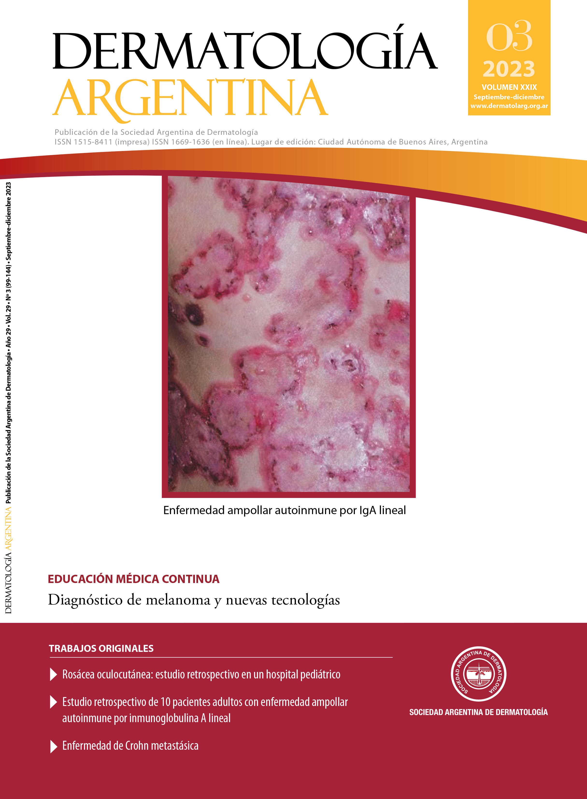
Melanoma diagnosis and new technologies
DOI:
https://doi.org/10.47196/da.v29i3.2483Keywords:
melanoma, diagnostic, molecular testing, immunohistochemistry, CGH, FISH, EGIRAbstract
It is known that all tumor lesions, including melanocytic ones, begin with varied genetic alterations that increase in number and characteristics from benignity to malignity.
In recent years, and based on these defined genetic alterations, other diagnostic methods have been developed. These new molecular tests would be useful in that limited number of lesions where histology and conventional immunohistochemistry do not distinguish benignity from malignity, and so we could be treating more or less a melanocytic lesion.
With this combination of a more accurate and early diagnosis and targeted white-specific treatments, we will be able to a better prognosis for patients with melanoma. The aim is to make these new molecular studies clinically useful.
References
I. Garbe C, Bauer J. Capítulo 113: Melanoma. En: Bolognia J, Schaffer J, Cerroni L. Dermatología 4ta edición. Elsevier España, Barcelona 2019;2000-2007.
II. Paek S, Sober A, Tsao H, Mihm M, Johnsoon T. Capítulo 124: Melanoma cutáneo. En: Wolff K, Goldsmith L, Katz S, Gilchrest B, Paller A, Leffell D. Fitzpatrick, Dermatology in General Medicine 7ma edición. Editorial Médica Panamericana, Buenos Aires 2009;1142-1145.
III. Conejo-Mir J, Moreno JC, Camacho F. Melanoma. Tratado de Dermatología. Editorial Océano, Barcelona 2012;1214-1216.
IV. Elder D, Elenitsas R, Murphy G, Xu X. Chapter 28: Benign pigmented lesions and malignant melanoma. En: Elder DE, Elenitsas R, Murphy GE, et ál., editor. Lever’s, histopathology of the skin. 10th edition. Philadelphia. Lippincott Williams & Wilkins;2009:744-759.
V. Gershenwald J, Scolyer R, Hess K, Sondak V, et ál. Melanoma staging: evidence-based changes in the American Joint Committee on Cancer eighth edition cancer staging manual. CA Cancer J Clin. 2017;67:472-492.
VI. Parigini AM, de Diego MC, Valdez R, Busso C. Anásisis de las modificaciones en la estadificación del T según el manual del American Joint Committee con Cancer. ¿Cómo cambia nuestro manejo de los pacientes? Estudio de corte transversal. Dermatol Argent. 2020;26:23-25.
VII. Fuentes L, Santonja C, Kutzner H, Requena L. Inmunohistoquímica en dermatopatología: revisión de los anticuerpos utilizados con mayor frecuencia (parte I). Actas Dermosifiliogr. 2013;104:99-127.
VIII. Saleem A, Narala S, Raghavan SS. Immunohistochemistry in melanocytic lesions: Updates with a practical review for pathologists. Seminars in Diagnostic Pathology. 2022;39:239-247.
IX. March J, Hand M, Truong A, Grossman D. Practical application of new technologies for melanoma diagnosis: Part II. Molecular approaches. J Am Acad Dermatol. 2015;72:943-958.
X. Andrea AA. Molecular testing for melanocytic tumors: a practical update. Histopathology. 2022;80:150-165.
XI. Fernández-Flores A. Conceptos modernos en tumores melanocíticos. Actas Dermosifiliogr. 2023;114: 402-412.
XII. Ferrara G, De Vanna AC. Fluorescence in situ hybridization for melanoma diagnosis: a review and a reappraisal. Am J Dermatopathol. 2016;38:253-269.
XIII. Nedergaard-Thomsen IM, Heerfordt IM, Karmisholt KE, Mogensen M. Detection of cutaneous malignant melanoma by tape stripping of pigmented skin lesions. A systematic review. Skin Res Technol. 2023;29(3):e13286.
Downloads
Published
Issue
Section
License
Copyright (c) 2023 on behalf of the authors. Reproduction rights: Argentine Society of Dermatology

This work is licensed under a Creative Commons Attribution-NonCommercial-NoDerivatives 4.0 International License.
El/los autor/es tranfieren todos los derechos de autor del manuscrito arriba mencionado a Dermatología Argentina en el caso de que el trabajo sea publicado. El/los autor/es declaran que el artículo es original, que no infringe ningún derecho de propiedad intelectual u otros derechos de terceros, que no se encuentra bajo consideración de otra revista y que no ha sido previamente publicado.
Le solicitamos haga click aquí para imprimir, firmar y enviar por correo postal la transferencia de los derechos de autor


