


Retrospective study of 10 adult patients with linear IgA autoimmune bullous disease
DOI:
https://doi.org/10.47196/da.v29i3.2485Keywords:
linear IgA bullous dermatosis, dapsone, autoimmune bullous diseasesAbstract
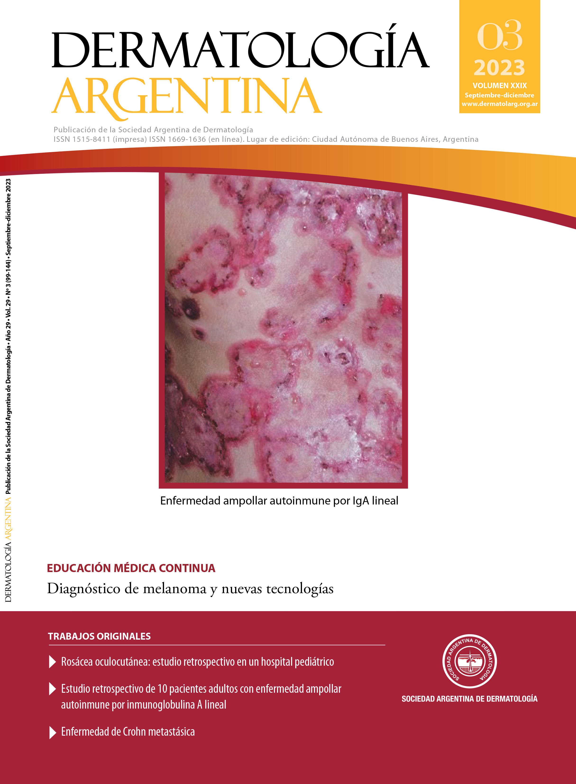
Background: linear IgA autoimmune blistering disease (LABD) is a rare, chronic and acquired autoimmune disease that presents with subepidermal blisters with IgA deposits in a linear pattern along in the basement membrane zone (BMZ) observed by direct immunofluorescence or immunohistochemical techniques.
Objectives: the aim of this study was to determine the epidemiological and clinical characteristics and the response to treatment of patients with LABD.
Design: a descriptive, observational and retrospective study was performed.
Materials and methods: 648 clinical histories were evaluated, including 10 patients with a confirmed diagnosis of LABD who attended the autoimmune bullous diseases section of the dermatology service of the José María Ramos Mejía Acute General Hospital between November 1997 and December 2021.
Results: of 648 patients, 10 were diagnosed with LABD (1.54% of all patients). The most affected sex was female. Mean age at diagnosis was 52.5 years. Only 20% presented mucosal involvement. All patients presented in the direct immunofluorescence (DIF) linear deposit of IgA in the BMZ. 80% were treated with dapsone associated with methylprednisone.
Conclusions: we highlight that the symptoms are heterogeneous, so the entity is defined by the finding in the DIF of IgA deposits with a linear pattern following the ZMB. In this study, unlike the literature, the age of onset was younger, mucosal involvement less frequent, and we observed emotional stress as a trigger not previously mentioned in the bibliography.
References
I. Chorzelski TP, Jablonska S. IgA linear dermatosis of childhood (chronic bullous disease of childhood). Br J Dermatol. 1979;101:535-542.
II. Hull CM, Zone JJ. Dermatitis herpetiforme y dermatosis ampollosa por Ig A lineal. En: Bolognia JL, Schaffer JV, Cerroni L (eds.). Dermatología. 4.ª ed. Barcelona: Elsevier, 2019:527-537.
III. Egan CA, Zone JJ. Linear IgA bullous dermatosis. Int J Dermatol. 1999;38:818-827.
IV. Díaz MS, Morita L, Ferrari B, Sartori S, et ál. Dermatosis ampollar IgA lineal: serie de 17 casos. Actas Dermosifiliogr. 2019;110:673-680.
V. Horiguchi Y, Ikoma A, Sakai R, Masatsugu A, et ál. Linear IgA dermatosis: report of an infantile case an analysis of 213 cases in Japan. J Dermatology. 2008;35:737-743.
VI. Di Milia M. Caso 24. Diagnóstico. Dermatosis por IG A lineal. En: Forero O, Candiz ME, Olivares L. Dermatosis ampollares autoinmunes. Haga su diagnóstico. 1.a ed. Argentina: Journal 2021:241-243.
VII. Genovese G, Venegoni L, Fanoni D, Muratori S, et ál. Linear IgA bullous dermatosis in adults and children: a clinical and immunopathological study of 38 patients. Orphanet J Rare Dis. 2019;14:115.
VIII. Tula M, Pazos M, Friedman P, Emilia N. Cohen Sabban, et ál. Dermatosis a IgA lineal asociada a fármacos: a propósito de un caso. Arch Argent Dermatol. 2015;65:45-49.
IX. Rodríguez L, Forero OL, Olivares L, Candiz ME, et ál. Dermatosis por IgA lineal vinculada a fármacos. Dermatol Argent. 2017;23:42-45.
X. Han J, Russo G, Stratman S, Psomadakis CE, et ál. Toxic epidermal necrolysis-like linear IgA bullous dermatosis after third Moderna COVID-19 vaccine in the setting of oral terbinafine. JAAD Case Rep. 2022;24:101-104.
XI. Fortuna G, Salas-Alanis JC, Guidetti E, Marinkovich MP. A critical reappraisal of the current data on drug-induced linear immunoglobulin A bullous dermatosis: A real and separate nosological entity? J Am Acad Dermatol. 2012;66:988-994.
XII. Chanal J, Ingen-Housz-Oro, Ortonne N, Duong TA, et ál. Linear Ig A bullous dermatosis: comparison between the drug-induced and spontaneous forms. Br J Dermatol. 2013;169:1041-1044.
XIII. Shipman AR, Reddy H, Wojnarowska F. Association between the subepidermal autoimmune blistering diseases linear IgA disease and the pemphigoid group and inflammatory bowel disease: two case reports and literature review. Clin Exp Dermatol. 2012;37:461-468.
XIV. Whittington CP, Huelsman MM, Bogner RR, Callen JP. IgA pemphigus and linear IgA bullous dermatosis in a patient with ulcerative colitis. Australas J Dermatol. 2020;61:e443-e445.
XV. Lings K, Bygum A. Linear IgA bullous dermatosis: a retrospective study of 23 patients in Denmark. Acta Derm Venereol. 2015;95:466-471.
XVI. Eguia del Valle A, Aguirre Urízar JM, Martínez Sahuquillo A. Manifestaciones orales de la enfermedad por depósito de Ig A lineal. Med Oralpatol Oral Ci Bucal. 2004;9:39-44.
XVII. Kashyap RR, Nair GR, Kashyap RS. Dilemmatic presentation of linear IgA disease: a case report. Stomatological Dis Sci. 2017;1:93-96.
XVIII. Ramos-Castellón C, Ortiz-Nieva G, Fresán F, Villalvazo L, et ál. Ocular involvement and blindness secondary to linear IgA dermatosis. J Ophthalmol. 2010. 2010:280396.
XIX. Zone JJ, Taylor TB, Kadunce DP, Meyer LJ. Identification of the cutaneous basement membrane zone antigen and isolation of antibody in linear immunoglobulin A bullous dermatosis. J Clin Invest. 1990;85:812-820.
XX. Matsuura K, Ujiie H, Hayashi M, Muramatsu K, et ál. Linear IgA bullous dermatosis in a pregnant woman with autoantibodies to the non-collagenous 16A domain of type XVII collagen. Acta Derm Venereol. 2017;97:404-405.
XXI. Horváth B, Niedermeier A, Podstawa E, Müller R, et ál. IgA autoantibodies in the pemphigoids and linear IgA bullous dermatosis. Exp Dermatol. 2010;19:648-653.
XXII. Kasperkiewicz M, Zillikens D, Schmidt E. Pemphigoid diseases: pathogenesis, diagnosis, and treatment. Autoimmunity. 2012;45:39-44.
XXIII. Izaki S, Mitsuya J, Okada T, Koga H, et ál. A Case of Linear IgA/IgG bullous dermatosis with anti-laminin-332 autoantibodies. Acta Derm Venereol. 2015;95:359-360.
XXIV. Hashimoto T, Ishii N, Tsuruta D. Production of neoepitopes by dynamic structural changes on BP180/type XVII collagen. J Invest Dermatol. 2017;137:2462-2464.
XXV. Shin L, Gardner JT 2nd, Dao H Jr. Updates in the diagnosis and management of linear IgA disease: a systematic review. Medicina (Kaunas). 2021;57:818.
XXVI. Pinard C, Hebert V, Lecuyer M, Sacre L, et ál. Linear IgA bullous dermatosis treated with rituximab. JAAD Case Rep. 2019;5:124-126.
XXVII. Benbenisty KM, Bowman PH, Davis LS. Localized linear Ig A disease responding to colchicine. Int J Dermatol. 2002;41:56-58.
XXVIII. He Y, Shimoda M, Ono Y, Villalobos IB et ál. Persistence of autoreactive IgA-secreting Bcells despite multiple inmunosuppressive medications including rituximab. JAMA Dermatology. 2015;151:646-650.
XXIX. Antiga E, Caproni M, Fabbri P. Linear immunoglobulin A bullous dermatosis: need for an agreement on diagnostic criteria. Dermatology. 2013;226:329-332.
XXX. Gottlieb J, Ingen-Housz-Oro S, Alexandre M, Grootenboer-Mignot S, et ál. Idiopathic linear IgA bullous dermatosis: prognostic factors based on a case series of 72 adults. Br J Dermatol. 2017;177:212-222.
Downloads
Published
Issue
Section
License
Copyright (c) 2023 on behalf of the authors. Reproduction rights: Argentine Society of Dermatology

This work is licensed under a Creative Commons Attribution-NonCommercial-NoDerivatives 4.0 International License.
El/los autor/es tranfieren todos los derechos de autor del manuscrito arriba mencionado a Dermatología Argentina en el caso de que el trabajo sea publicado. El/los autor/es declaran que el artículo es original, que no infringe ningún derecho de propiedad intelectual u otros derechos de terceros, que no se encuentra bajo consideración de otra revista y que no ha sido previamente publicado.
Le solicitamos haga click aquí para imprimir, firmar y enviar por correo postal la transferencia de los derechos de autor